Types of Hair
There are three types of hair. Thick, pigmented hairs are called terminal hairs. Terminal hairs on the top of the head and in the beard, axillary, and pubic areas are influenced by androgens. Androgens are important in regulating hair growth.
At puberty, androgens increase the size of follicles in the beard, chest, and limbs and decrease the size of follicles in the bitemporal region, which reshapes the hairline in men and many women. Lanugo hairs are the fi ne hairs found on the fetus; similar fi ne hairs (peach fuzz) found on the adult are called vellus hairs. Vellus hair is short, fi ne, and relatively nonpigmented and covers much of the body. Hair on the rest of the body is independent of androgens.
Hair Structure
The hair shaft is dead protein. It is formed by compact cells that are covered by a delicate cuticle composed of platelike scales. The living cells in the matrix multiply more rapidly than those in any other normal human tissue. They push up into the follicular canal, undergo dehydration, and form the hair shaft, which consists of a dense, hard mass of keratinized cells. Normal hairs have a pointed tip. The hair in the follicular canal forms a cylinder of uniform diameter. Short hairs with tapered tips either have short growth cycles or have experienced the recent onset of anagen. The growing shaft is surrounded by several concentric layers.
The outermost glycogen-rich layer is called the outer root sheath. It is static and continuous with the epidermis. The inner root sheath (Henle’s layer, Huxley’s layer, and cuticle) is visible as a gelatinous mass when the hair is plucked. It protects and molds the growing hair but disintegrates before reaching the surface at the infundibulum. The hair shaft that emerges has three layers—an outer cuticle, a cortex, and sometimes an inner medulla—all of which are composed of dead protein. The cuticle protects and holds the cortex cells together. Split ends result if the cuticle is damaged by brushing or chemical cosmetic treat
The cortex cells in the growing hair shaft rapidly synthesize and accumulate proteins while in the lower regions of the hair follicle. Systemic diseases and drugs may interfere with the metabolism of these cells and reduce the hair shaft diameter. Pigment-containing melanosomes are acquired deep in the bulb matrix and are deposited in the cortical and medullary cells.
Hair Follicle
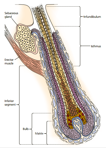
Humans have about 5 million hair follicles at birth. No follicles are formed after birth, but their size changes under the infl uence of androgens.The hair follicle is formed in the embryo by a club-shaped epidermal down-growth—the primary epithelial germ that is invaginated from below by a flame-shaped, capillarycontaining dermal structure called the papilla of the hair follicle. The central cells of the down-growth form the hair matrix, the cells of which form the hair shaft and its surrounding structures. The matrix lies deep within the subcutaneous fat. The mature follicle contains a hair shaft, two surrounding sheaths, and a germinative bulb
The follicle is divided into three sections. The infundibulum extends from the surface to the sebaceous gland duct. The isthmus extends from the duct down to the insertion of the erector muscle. The inferior segment, which exists only during the growing (anagen) phase, extends from the muscle insertion to the base of the matrix. The matrix contains the cells that proliferate to form the hair shaft The mitotic rate of the hair matrix is greater than that of any other organ. The cells begin to differentiate at the top of the bulb.
The inner and outer root sheaths protect and mold the growing hair. The inner root sheath disintegrates at the duct of the sebaceous gland. Hair growth is greatly influenced by any stress or disease process that can alter mitotic activity.